Case Presentation: CEREC Emax #11

August 7, 2013

contributed by Chandan Jaspal:

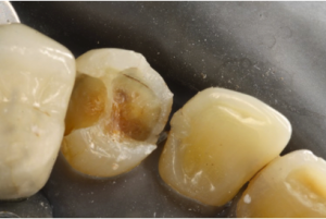
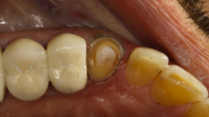
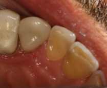
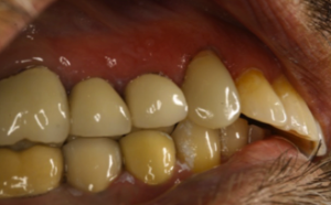
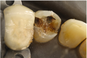
Summary – Tooth # 11 had large composite fillings with recurrent decay. The patient came with pieces of the composite in his hand. After doing the COAD (Clean Out And DIagnose and determining the restorability, we decided to restore the tooth with an Emax Crown using the CEREC CAD/CAM System.  Our rationale for using Emax was due to the lack of interarch space due to wear.  Our team felt that using a high strength monolithic material would not only provide better esthetics, but better use the space offered by the occlusal and axial reduction.  To match this new crown with the natural looking canine on the other side of the arch (tooth #6), we utilized  “Biogeneric reference” on the CEREC machine.  The biogeneric reference function asked us if we wanted to mirror the contralateral tooth. Tooth #6 had a flat incisal surface, and the crown proposed by the CEREC came out with a normal architecture “triangular” shape.  Minor modifications were made, but the patient wound up loving the computer’s design better than his existing tooth both in apperance and feel.  It did raise an issue however that the proposal using biogeneric reference did NOT really resemble the tooth we wished to copy.  This is probably why Biogeneric Copy using a diagnostic wax-up is still the preferred method to use for CEREC crowns made in the anterior region.

Decayed Tooth

Decayed Canine

cleanout

Tooth structure remaining after COAD procedure.  Tooth remained Asymptomatic after Fuji Lining LC and definitive Core Flow build up was placed.

prep

Prep

finalocclusal

Final Occlusal View

finalbuccal

Final Buccal view after occlusion was checked.

Crown Lengthening

June 5, 2013

contributed by:  Chandan Jaspal

INDICATIONS: (2 types, Restorative and Aesthetic)

Restorative/functional crown lengthening Indications

  • To access to subgingival caries
  • To increase the clinical crown height reduced by tooth wear, caries or a
  • fracture extending subgingivally
  • To assist in creating a ‘ferrule’ effect
  • Correcting the position of the restorative margin when there has been invasion
  • of the biologic width
  • Access to superficial root perforations (e.g. following pin placement)

Aesthetic crown lengthening Indications

  • Correction of short clinical crowns due to wear or altered passive eruption
  • Creating gingival symmetry in the smile line
  • Correcting irregular/uneven gingival margins
  • Correcting for excessive gingival “Gummy smile” or hyperplasic tissue overgrowth

Contraindications to Crown Lengthening

  •  Smoking is a contra-indication to most gum surgeries including crown lengthening.
  •  Thin gum tissue
  •  Poor oral hygiene
  •  Where the furcation between the roots of a molar tooth will become exposed following crown lengthening.
  •  Unfavorable crown to root ration being created by crown lengthening.  I.E. The crown becoming longer than the root following crown lengthening surgery.
  •  Front teeth with long clinical crowns.
  •  An un-restorable tooth after surgery

 ALTERNATIVES TO CROWN LENGTHENING: 

  • Invisalign or Orthodontic Bracket tooth extrusion
  • Extraction of the tooth and replacing it with a dental Implant or a dental bridge.
(Photos to follow)

Crossbite

March 19, 2012

contributed by Anahita Gupta:

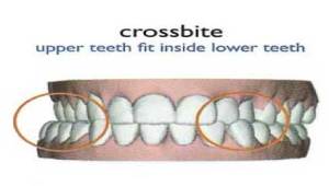
A crossbite is an irregularity of the occlusal surface of the tooth. It occurs when one or more teeth have a buccal (Mandibular teeth) or lingual (Maxillary teeth) position when compared to its matching tooth above or below it.

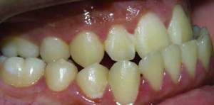
Crossbite Example

The crossbite can be:-

  1. “Anterior”, with a negative overjet, also known as a class III skeletal abnormality or prognathism.
  2. “Posterior” crossbite, with a narrow maxillary bone and a narrow upper dental arch.
  3. Crossbite can also be either unilateral or bilateral.

Crossbite

Etiology:

  1. 1.     Hereditary/Genetic- This usually presents as a wide lower jaw thus affecting the entire dentition.
  2. 2.     Delayed loss of Deciduous teeth- This usually presents as a single tooth cross.
  3. 3.     Abnormal path of eruption
  4. 4.     Thumb sucking can contribute to a crossbite by constricting the width of the palate and deforming the upper bone of the palate.


Symptoms:


  1. Painful chewing and a painful jaw and teeth.
  2.  It can also affect chewing in such a way that the temporomandibular joint gets inflamed. This inflammation causes pain in the TMJ and sometimes headaches.


Complications:

  1. Periodontitis and gingivitis
  2. Tooth loss due to malocclusion
  3. Degeneration of the temporomandibular joint, causing arthritis of the TMJ and wearing away of the joint surface. This can result in the need for surgery to replace the TMJ completely.


Crossbite prevention:

  1. 1.     
You can prevent a crossbite by stopping thumb sucking early in life so the upper palate doesn’t become narrowed and malformed.
  2. 2.     You can have the deciduous teeth extracted when the permanent are about to erupt.
  3. 3.     Many types of crossbite, however, cannot be prevented and must be treated when noted.


Treatment options:

Orthodontists need to treat a crossbite as soon as possible so that the teeth can be realigned. If mouth breathing is part of the cause, a tonsillectomy and adenoidectomy should be performed before the patient undergoes orthodontic therapy.

I) The first part of treatment involves maxillary expansion. It widens the jaw when worn nightly for a couple of months. A key turns the device and gradually increases the width of the palate.

II) After the palate has expanded enough, orthodontic devices are necessary to straighten the teeth. This means a full set of braces for a total of one to two years in order to create an ideal bite.


Tooth Bleaching

October 3, 2011

contributed by: Shahed Al Khalifah

Tooth discoloration usually occurs due to patient- or dentist-related causes:

BLEACHING MATERIALS

Hydrogen Peroxide. The 30 to 35% stabilized aqueous solutions are the most common.

Sodium Perborate. This oxidizing agent is available in a powdered form or as various commercial preparations. It contains about 95% perborate, corresponding to 9.9% of the available oxygen.

Carbamide Peroxide. This agent, also known as urea hydrogen peroxide, is available in the concentration range of 3 to 45%. The popular commercial preparations contain about 10% carbamide peroxide with approximately 3.5% hydrogen peroxide.

BLEACHING MECHANISM

Bleaching agents, mainly oxidizers, act on the organic structure of the dental hard tissues, slowly degrading them into chemical by-products, such as carbon dioxides, that are lighter in color. The oxidation-reduction reaction that occurs during bleaching is known as a redox reaction.

BLEACHING TECHNIQUES

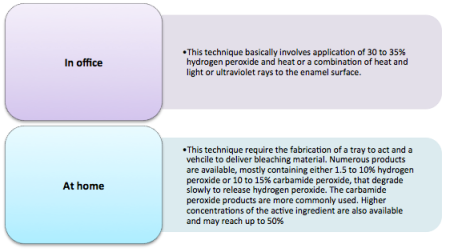
External bleaching. In these techniques, oxidizers are applied to the external enamel surface of the teeth. Mainly used in case of vital teeth.

In Office or At Home Bleaching

Contraindications for external bleaching:

  • Severe enamel loss.
  • Hypersensitive teeth.
  • Defective coronal restorations.
  • Presence of caries.
  • Allergy to bleaching gels.
  • Bruxism.


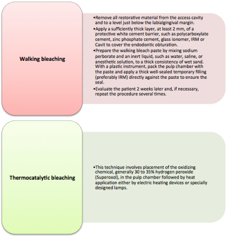
Internal bleaching. Used for endodontically treated teeth. It may be successfully carried out many years after root canal therapy and discoloration.

Walking bleaching and Thermocatalytic bleaching

Contraindications for internal bleaching:

  • Superficial enamel discoloration.
  • Defective enamel formation.
  • Severe dentin loss.
  • Presence of caries.
  • Discolored composites.